234. What is Hoover’s sign ?
*. Hoover’s sign is a clinical sign used to differentiate between unilateral organic and functional leg weakness
*. This is based on the principle ofsynkinetic movements
Eliciting Hoover’s sign
*. Patient is asked to lie down in supine position
*. The examiner keeps the palm ofhis hand under the heel of the patient’s normal leg
*. The patient is asked to lift his ‘paretic’ leg
*. Normally there is synkinetic hip extension in the normal limb and the examiner can feel the pressure on his palm
*. In patients with organic paresis, there is accentuation ofthis response and the examinercan use the straightened normal limb as a lever to lift the whole limb and lower body
*. In patients with functional paresis, there is no synkinetic hip extension and the examinerfails to appreciate any downward force
*. This occurs because the patientdoes not make any effort to raise the leg
*. Similarly, the patient can be asked to raise the normal limb
*. Patients with functional leg paresis unconsciously press down with the ‘paretic’ limb, whereas no response is obtained in those with organic paresis
233. What is Buerger's disease ?
Buerger’s disease, also called thromboangitis obliterans, is a rare form of vasculitis characterized by acute inflammation and clotting of the arteries and veins of the hands and feet. The inflammation can lead to blockages of the arteries of the lower portions of the armsand legs. These blockages can cause critical limb ischemia and/or claudication , rest pain and non-healing sores or ulcers, in the hands and/or feet.
Buerger’s disease, which occurs almost exclusively in heavy smokers, is different from peripheral arterial disease (PAD) in that it is not caused by atherosclerosis (plaque buildup in the arteries). The disease can cause pain in the hands and feet in response to exercise due to reduced blood-flow.
232. What is a Felon ?
A purulent infection in the tight fascial plane adjacent to the terminal intraphalangeal joint of the fingers or toes, due to an open wound; as the inflammatory mass expands within the confined space, the vascular supply is compromised, predisposing the site to osteomyelitis, pulp necrosis and sloughing of tissue; the pain is very intense and seemingly disproportionate with the scant amount of swelling and erythema clinically evident.
231. What is a ganglion cyst ?
230. What is Marjolin's Ulcer ?
Marjolin's ulcer refers to an aggressive ulcerating squamous cell carcinoma presenting in an area of previously traumatized ,chronically inflamed, or scarred skin. They are commonly present in the context of chronic wounds including burn injuries , venous ulcers , ulcers from osteomyelitis, and post radiotherapy scars.
The term was named after French surgeon , Jean-Nicolas Marjolin , who first described the condition in 1828. The term was later coined by J C De Costa .
229. What is Trousseau sign ?
Trousseau sign of latent tetany is a medical sign observed in patients with low calcium. This sign may become positive before other gross manifestations of hypocalcemia such as hyperreflexia and tetany , but is generally believed to be more sensitive (94%) than the Chvostek sign (29%) for hypocalcemia.
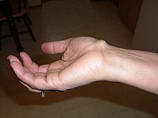
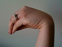
To elicit the sign, a blood pressure cuff is placed around the arm and inflated to a pressure greater than the systolic blood pressure and heldin place for 3 minutes. This will occlude the brachial artery . In the absence of blood flow, the patient's hypocalcemia and subsequent neuromuscular irritability will induce spasm of the muscles of the hand and forearm. The wrist and metacarpophalangeal joints flex,the DIP and PIP joints extend, and the fingers adduct . The sign is also known as main d'accoucheur.
228. What is Freiberg's infraction ?
*Freiberg's infraction* is a condition specific to the lesser metatarsal heads, most commonly the second metatarsal. Also known as avascular necrosis, or AVN, this condition results in a focal loss of blood supply to the metatarsal head. Freiberg's infraction occurs at the metaphysis of the metatarsal bone. The metaphysis in the region where the primary or nutrient artery of the bone supplies the distal metatarsal. Loss of blood supply to the metaphysis (infraction) results in collapse of the metatarsal head.
227. What is Roberts syndrome ?
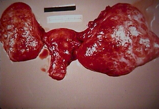
*Roberts syndrome* is a genetic disorder characterized by limb and facial abnormalities.They have shortened arm and leg bones (hypomelia), particularly the bones in their forearms and lower legs. In severe cases, the limbs may be so short that the hands and feet are located very close to the body (phocomelia).Individuals with Roberts syndrome typically have numerous facial abnormalities, including a cleft lip with or without cleft palate, micrognathia, ear abnormalities, wide-set eyes (hypertelorism), outer corners of the eyes that point downward (down-slanting palpebral fissures), small nostrils, and a beakednose. They may have microcephaly, and in severe cases encephalocele. In addition, people with Roberts syndrome may have heart, kidney, and genital abnormalities.
226. What is Fox's sign ?
225. What is Pancoasts tumor ?
224. What is Mazzotti reaction ?
223. What is Pallister-Hall syndrome ?
Pallister-Hall syndrome is a disorder that affects the development of many parts of the body. Most people with this condition have extra fingersand/or toes (polydactyly), and the skin between some fingers or toes may be fused (cutaneous syndactyly).An abnormal growth in the brain called a hypothalamic hamartoma is characteristic of this disorder. In many cases, these growths do not cause any medical problems; however, some hypothalamic hamartomas lead to seizures or hormone abnormalities that can be life-threatening in infancy. Other features of Pallister-Hall syndrome include a malformation of the airwaycalled a bifid epiglottis, an obstruction of the anal opening (imperforate anus), and kidney abnormalities. Although the signs and symptoms of this disorder vary from mild to severe, only a small percentage of affected people have serious complications.
Ans.222 *Krukenbergs tumor*
Ans.221 *May-Thurner syndrome*
Ans.220 *May-Hegglin anomaly*
*May-Hegglin anomaly* This is an autosomal dominant disorder associated with the presence of giant platelets, moderate thrombocytopenia and bright blue Döhle-body like inclusions in WBC that are composed of parallel filaments. Individuals with this anomaly are at risk for bruising, menorrhagia and complicated childbirth but 40% are asymptomatic.
Ans.219 *Hollenhorst Plaques*
Hollenhorst Plaques
*. Cholesterol emboli which usually dislodged from the carotid arteries, or calcific fragments from a stenosed aortic valve.
*. Clinical features:
*. Symptoms: usually give no symptoms.
*. Signs:
*. Planar or flat-shaped, minute, bright, refractile, golden-yellow crystals that most commonly found at bifurcations of retinal arteries
*. Blood flow may not be disrupted because of the shape of the crystal.
Ans.218 *Riglers sign*
*Riglers sign* , which is also known as the double-wall sign or the bas-relief sign , both sides of the bowel wall can be visualized on a radiograph of the abdomen obtained with the patient in the supine position.Gas normally outlines only the luminal surface of the bowel wall and not the serosal surface, which has a degree of opacity similar to that of adjacent peritoneal contents. When at least a moderate amount offree intraperitoneal air exists, however, this free air is more likely to accumulate between bowel loops,thus permitting visualization of the outer walls of the bowel.
Ans.217 *Maurers dots*
Ans.216 *Filatov-Dukes disease*
*Filatov-Dukes disease* , also known as Staphylococcal scarlet fever, caused by Staph. aureus
epidermolytic toxin, described in 1900.
It is characterized by:
*. Sore throat
*. Fever
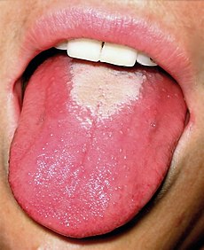
*. Bright red tongue with a"strawberry" appearance
*. Characteristic rash :
fine, red, and rough-textured; it blanches upon pressure.
*. appears 12–72 hours after the fever.
*. generally starts on the chest, armpits, and behind the ears.
*. spares the face (although some circumoral pallor is characteristic).
*. is worse in the skin folds . These Pastia lines (where the rash runs together in the armpits and groin) appear and can persist after the rash is gone.
*. may spread to cover the uvula .
*. The rash begins to fade three to four days after onset and desquamation (peeling) begins.
Ans.215 *Fournier's gangrene*
*Fournier's gangrene* is a type of necrotizing infection or gangrene usually affecting the perineum .
It was first described by Baurienne in1764 and is named after a French venereologist , Jean-Alfred Fournier following five cases he presented in clinical lectures in 1883. In the majority of cases Fournier's gangrene is a mixed infection causedby both aerobic and anaerobic bacteria.
Ans.214 *Stevens-Johnson syndrome*
Ans.213 *Hoffmans syndrome*
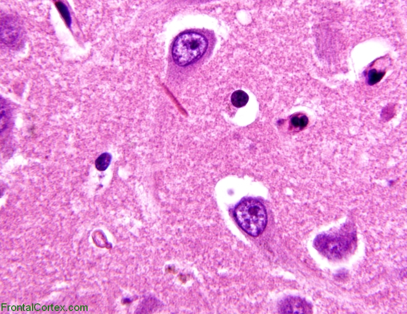
Ans.212 *Masson body*
*Masson body*
Granulation tissue plug found in lumen of small airways in bronchiolitis obliterans obstructive
pneumonia (BOOP).
Masson body was termed by Masson Riopelle and Martin in 1937.
Bronchiolitis obliterans organizing pneumonia ( BOOP ) is an inflammation of the bronchioles ( bronchiolitis )and surrounding tissue in the lungs . It is an non infectious pneumonia .BOOP is often caused by a pre-existing chronic inflammatory disease like rheumatoid arthritis . BOOP can also be a side effect of certain medicinal drugs, e.g. amiodarone . BOOP was discovered by Dr. Gary Epler in 1985.
Ans.211 *Cushing's ulcer*
Ans.210 *Martorell's ulcer*
In the 1940s, Martorell described a unique, ischemic ulcer of the leg as a complication of hypertension. Hines and Farber subsequently confirmed the existence of this ulcer and reported 11 cases of Martorell's ulcers. The classic presentation of this ulcer includes a superficial ulcer on the lower extremity, poorly controlled or long-standing diastolic arterial hypertension in both the extremities, bilateral symmetry that may manifest as a pigmented lesion on the contralateral leg, a history of minimal trauma, which may not be remembered, and moderate to severe pain often out of proportion to the size of the ulcer. The lesion is more common in female patients than in male patients, rarely seen in African Americans, and is most often seen in women between the ages of 55 and 65.These lesions are refractory to local wound care or more aggressive therapy and may progress by irregular extension of the ischemic area.
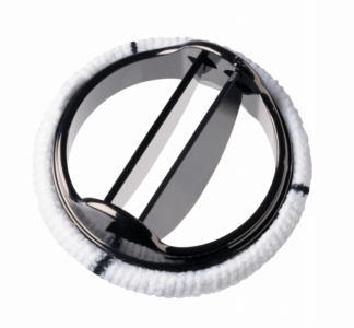
Ans.209 *St. Jude valve*
Ans. 208 *Barraquer-Simons Syndrome*
*Barraquer-Simons syndrome*
Acquired partial lipodystrophy; presents usually around 8-10, preceded generally by an acute viral
syndrome
infection; spares legs and hips; 1/3 of patients develop membranoproliferative glomerulonephritis;
associated with accelerated complement activation and a serum immunoglobulin G, called C3
nephritic factor that are felt to cause lysis of adipose tissue.It is named for Luis Barraquer Roviralta and Arthur Simons.
Figure 1.
Facial appearance of patient at 12 years of age showing loss of buccal fat, neck, trunk, and upper extremities' subcutaneous fat.
Figure 2. Facial appearance of patient at 6 years of age.
Figure 3.
Immunofluorescence micrograph from skin biopsy demonstrating granular accumulation of C3 along the basement membrane.
Ans.207 *Spurling's test*
Ans.206 *Riddoch’s sign*
*Riddoch’s sign*
In cerebellar disease, with outstretched hands, the hand on the affected side begins to hyperpronate,
so that the palm faces outward, and rises above the level of the other hand.
In cerebellar disease, with outstretched hands, the hand on the affected side begins to hyperpronate,
so that the palm faces outward, and rises above the level of the other hand.
Ans.205 *Sprengel scapula*
Ans.204 *Pallas sign*
Ans.203 *Barré-Liéou syndrome*
*Barré-Lieou syndrome* was discovered by and named after Jean Alexandre Barre, M.D., a French neurologist. Cervicosympathetic syndrome, characterized by neck pain and dizziness due to arthritic or
traumatic damage to the cervical spine.
Ans.202 *Hirschsprungs disease*
*Hirschsprungs disease*
Congenital aganglionic megacolon; colonic dilatation proximal to an aganglionic, contracted distal
colon and rectum; caused by gestational failure of neural crest cells to migrate to distal colon.
Autosomal dominant form associated with mutations of the RET gene, autosomal recessive form
associated with mutations of the endothelin-B-receptor gene.
Ans.201 *Barlow's maneuver*
Ans.200 *Curschmann's spirals*
Ans.199 *Currarino-Silverman syndrome*
Ans.198 *Hirata disease*
*Hirata disease* Hirata et al. first reported a patient with insulin autoimmune syndrome in 1970. Insulin autoimmune syndrome with hypoglycemia associated with insulin autoantibodies. Third
leading cause of spontaneous hypoglycemia in Japan; rare in Western countries.
Ans.197 *Achenbach's syndrome*
Ans.196 *Hirano bodies*
Ans.195 *Weill-Marchesani syndrome*
*Weill-Marchesani
syndrome*
Rare, autosomal recessive syndrome of ectopia lentis, short stature, brachydactyly, and glaucoma.
Associated with mutation in ADAMTS10 gene.
syndrome*
Rare, autosomal recessive syndrome of ectopia lentis, short stature, brachydactyly, and glaucoma.
Associated with mutation in ADAMTS10 gene.
Ans.194 *Currarino triad*
*Currarino triad*
Partial sacral agenesis with intact first sacral vertebra resembling a sickle; a presacral mass, and
anorectal malformation; associated with a mutation in a homeobox gene, HLXB9.
Partial sacral agenesis with intact first sacral vertebra resembling a sickle; a presacral mass, and
anorectal malformation; associated with a mutation in a homeobox gene, HLXB9.
Ans.193
*Barlow's disease*
>Infantile scurvy (named after Sir Thomas Barlow (1845–1945)).
>Mitral valve prolapse
Ans.193
*Barlow's disease*
>Infantile scurvy (named after Sir Thomas Barlow (1845–1945)).
>Mitral valve prolapse
>Infantile scurvy (named after Sir Thomas Barlow (1845–1945)).
>Mitral valve prolapse
Ans.192 *Abernethy malformation*
*Abernethy malformation* Congenital anomaly of the splanchnic vasculature, arises from defects in vitelline vein formation.
#Type 1, portal vein completely diverted into IVC and complete absence of formation of intrahepatic
portal vein.
#Type 2, portal venous system is formed, but there is an abnormal communication with
systemic veins, usually IVC; associated with hepatic encephalopathy and hepatopulmonary
syndrome.
#Type 1, portal vein completely diverted into IVC and complete absence of formation of intrahepatic
portal vein.
#Type 2, portal venous system is formed, but there is an abnormal communication with
systemic veins, usually IVC; associated with hepatic encephalopathy and hepatopulmonary
syndrome.
Ans.191 *Fothergill's sign*
*Fothergill's sign*
In rectus sheath hematomas, tender mass that does not cross the midline and remains palpable when
patient tenses the rectus muscle (bluish discoloration not usually seen until 3 or 4 days).
In rectus sheath hematomas, tender mass that does not cross the midline and remains palpable when
patient tenses the rectus muscle (bluish discoloration not usually seen until 3 or 4 days).
Ans.190 *Weil-Felix reaction*
Weil-Felix reaction
Proteus cell wall O antigens, such as OX-2, OX-19, and OX-K, cross-reacting with antigens of
several species of rickettsiae.
Proteus cell wall O antigens, such as OX-2, OX-19, and OX-K, cross-reacting with antigens of
several species of rickettsiae.
Subscribe to:
Posts (Atom)